


 收藏产品
收藏产品 产品信息
- Genetically modified cell lines best reflect MOA (Mechanism of Action)
- Higher activity and larger assay window for robust and reproducible cell-based bioassay
- Comprehensive application data to support assay development and validation
- Full tracible record, stringent quality control and validated cell passage stability
- Parental cell line legally obtained from internationally recognized cell resource bank and commercially licensed
- Global commercial license assistance whenever regulatory filing is required
描述(Description)
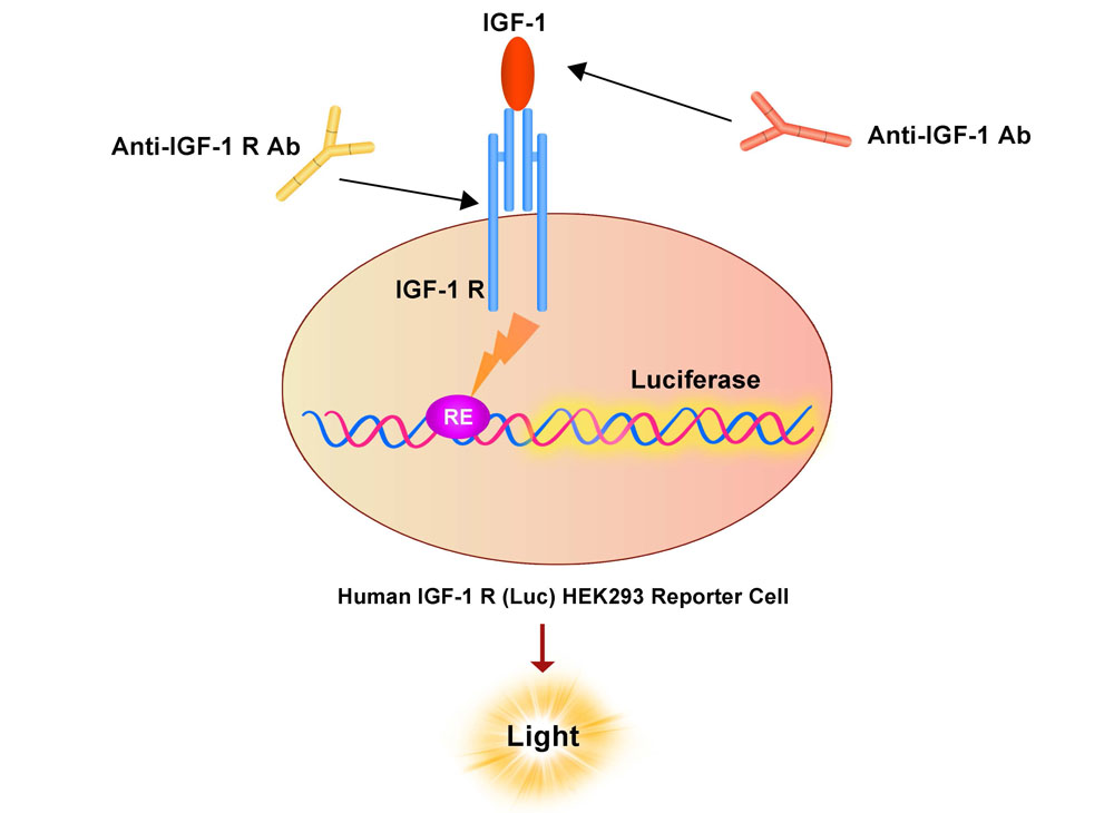
The Human IGF-1 R (Luc) HEK293 Reporter Cell was engineered to not only express signaling response element, but also express the receptor full length human IGF-1 R (Gene ID: 3480). When stimulated with human IGF-1 protein, the IGF-1/IGF-1 R interaction drives RE-mediated luminescence. Neutralization of biological effect of human IGF-1 protein by corresponding antibody results in a decrease in luminescence.
应用说明(Application)
• Screen for neutralizing antibodies blocking the stimulation of human IGF-1 protein.
生长特性(Growth Properties)
Adherent
筛选标记(Selection Marker)
Puromycin (2 μg/mL) + Hygromycin (20 μg/mL)
培养基(Complete Growth Medium)
DMEM medium + 10% FBS
冻存液(Freeze Medium)
Serum-free cell cryopreservation medium
装量(Quantity)
1 vial contains at least 5×10^6 cells in 1 mL serum-free cryopreservation medium
存储(Storage)
Frozen in liquid nitrogen.
支原体检测(Mycoplasma Testing)
Negative
无菌检测(Sterility Testing)
Negative
使用说明(Instructions for Use)
See data sheet for detailed culturing and assay protocol.
产品数据图
Receptor Assay
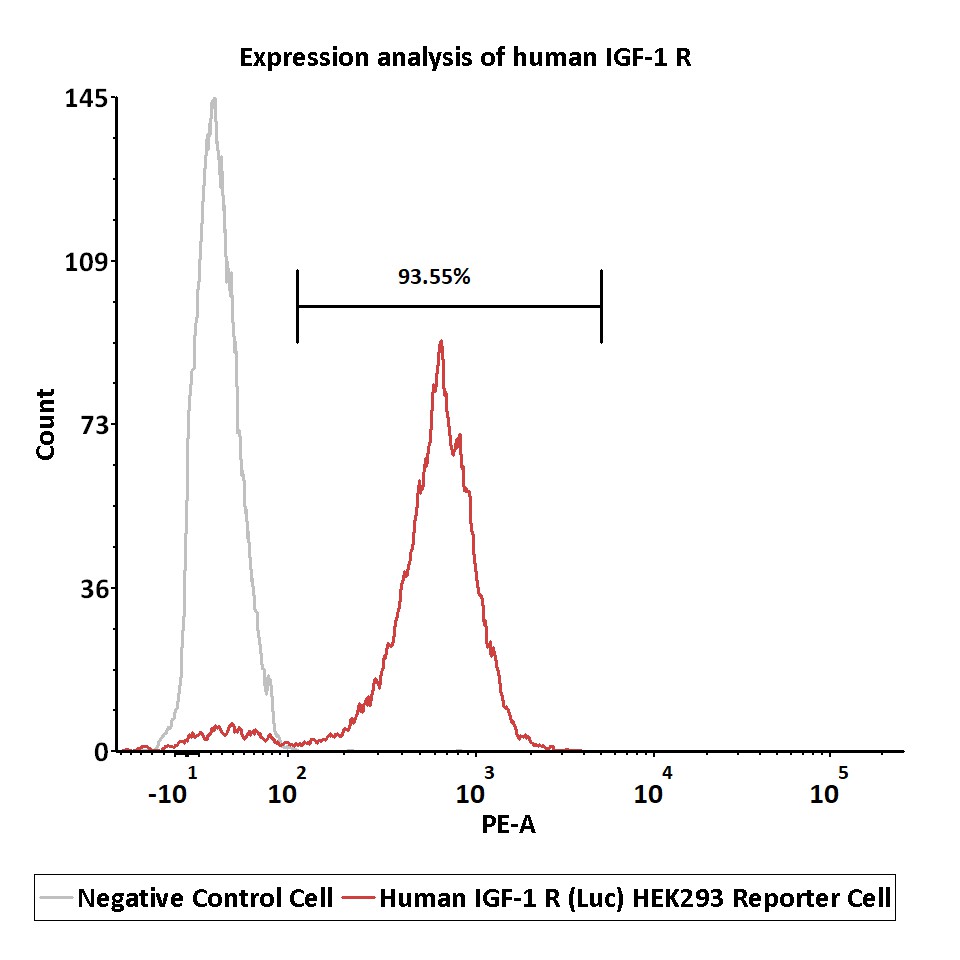
Expression analysis of human IGF-1 R on Human IGF-1 R (Luc) HEK293 Reporter Cell by FACS.
Cell surface staining was performed on Human IGF-1 R (Luc) HEK293 Reporter Cell or negative control cell using PE-labeled anti-human IGF-1 R antibody.
Protocol
Application
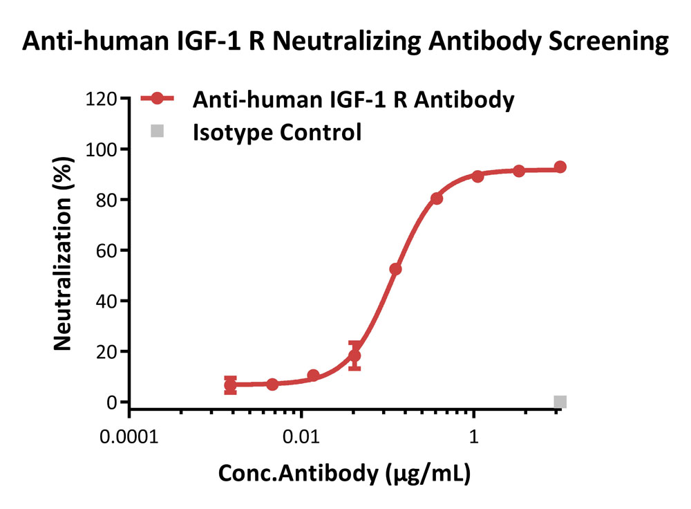
Inhibition of human IGF-1 protein-induced reporter activity.
This reporter cell was incubated with serial dilutions of antibodies in the presence of human IGF-1 protein (Cat. No. IG1-H5245) with a final concentration of 0.1 μg/mL. The EC50 of anti-human IGF-1 R neutralizing antibody (Teprotumumab) is approximately 0.12 μg/mL.
Protocol
Signaling Bioassay
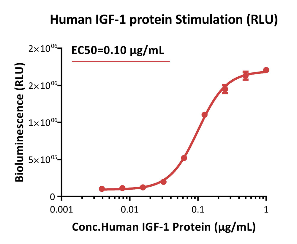
Response to human IGF-1 protein (RLU).
This reporter cell was incubated with serial dilutions of human IGF-1 protein (Cat. No. IG1-H5245). The EC50 was approximately 0.10 μg/mL.
Protocol
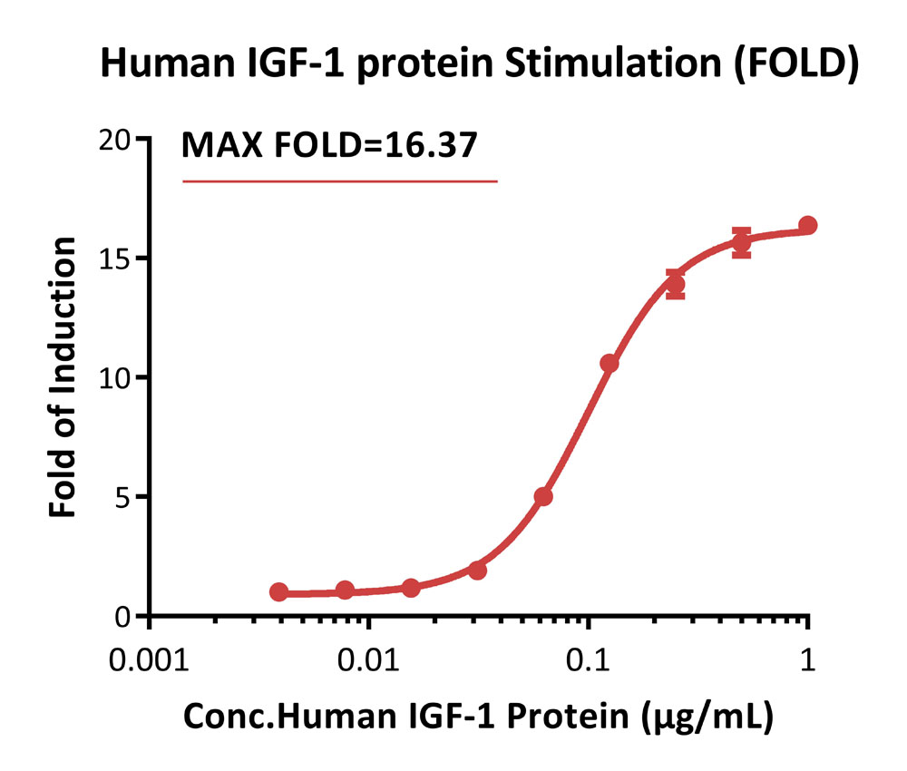
Response to human IGF-1 protein (FOLD).
This reporter cell was incubated with serial dilutions of human IGF-1 protein (Cat. No. IG1-H5245). The max induction fold was approximately 16.37.
Protocol
Passage Stability
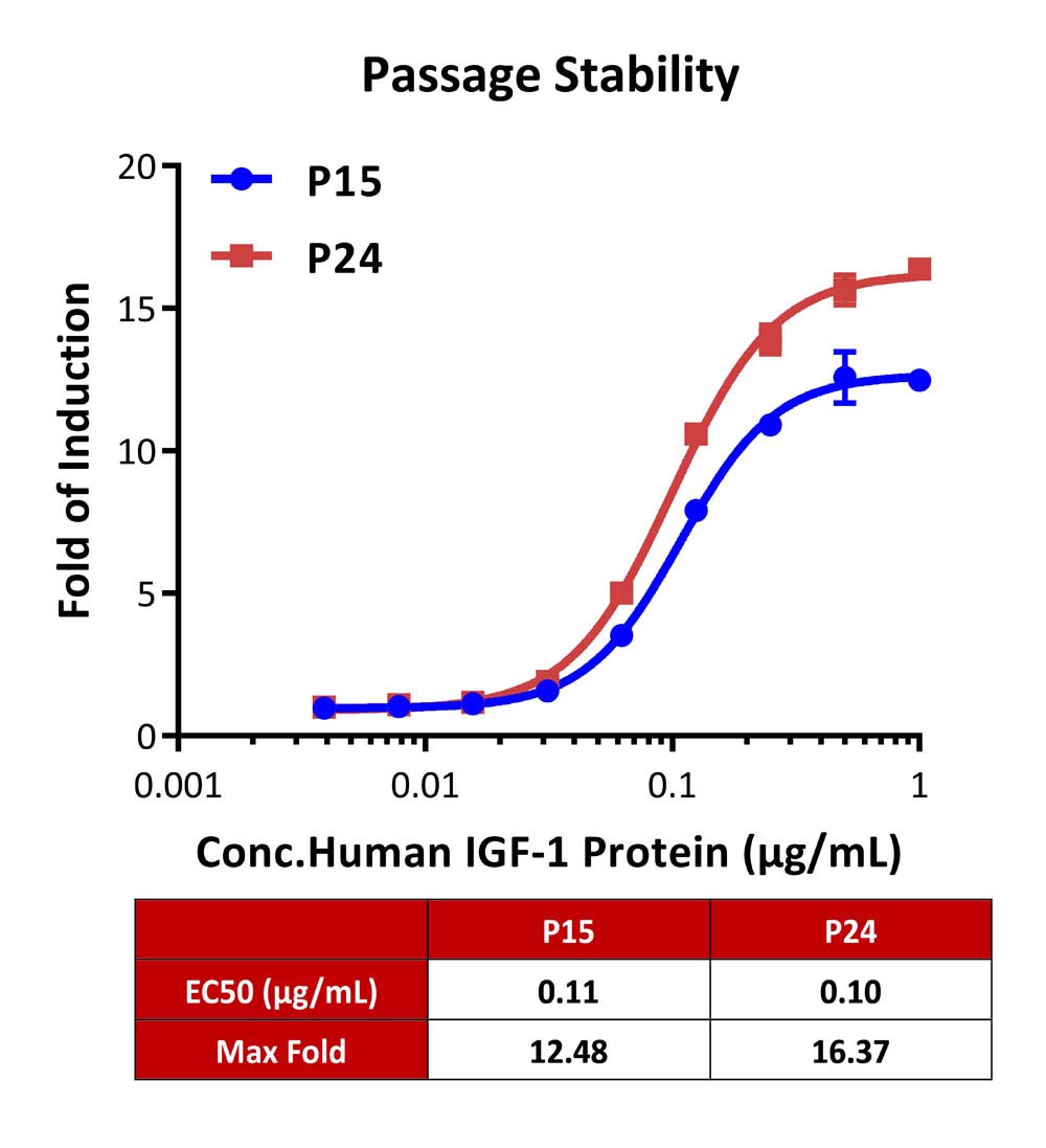
Passage stability analysis by Signaling Bioassay.
The continuously growing Human IGF-1 R (Luc) HEK293 Reporter Cell was stimulated with serial dilutions of human IGF-1 protein (Cat. No. IG1-H5245). Human IGF-1 protein stimulated response demonstrates passage stabilization (fold induction and EC50) across passage 15-24.
Protocol
产品评论 发表评论

背景
The Insulin-like Growth Factor 1 Receptor (IGF1) is also known as CD221, JTK13. and is a transmembrane receptor that is activated by IGF-1 and by the related growth factor IGF-2. It belongs to the large class of tyrosine kinase receptors. This receptor mediates the effects of IGF-1, which is a polypeptide protein hormone similar in molecular structure to insulin. IGF1R is make up of two alpha subunits and two beta subunits ,the Both the α and β subunits are synthesized from a single mRNA precursor. The precursor is then glycosylated, proteolytically cleaved, and crosslinked by cysteine bonds to form a functional transmembrane αβ chain.The α chains are located extracellularly while the β subunit spans the membrane and are responsible for intracellular signal transduction upon ligand stimulation. IGF1R have a binding site for ATP, which is used to provide the phosphates for autophosphorylation. There is a 60% homology between IGF1R and the insulin receptor. In response to ligand binding, the α chains induce the tyrosine autophosphorylation of the β chains. This event triggers a cascade of intracellular signaling that, while somewhat cell type specific, often promotes cell survival and cell proliferation.
 靶点信息
靶点信息  数据表和文档
  联系我们
联系我们 


 项目合作
项目合作