- English
- 日本語
- 한국어
- Deutsch
- Français
- Español
No data
官方服务号
招聘公众号

ACROBiosystems百普赛斯基于流式细胞术平台,经严格的方法学验证,提供多样的流式分析方案,精准识别细胞组成及状态。我们提供多种荧光染料标记的流式抗体和预混试剂盒,满足您在流式细胞仪上的各种需求,为您在临床前药物开发、临床试验免疫监测等研究提供高质量、高效率的免疫细胞分型分析工具!
如有更多荧光染料标记或配色需求,请点击 咨询。
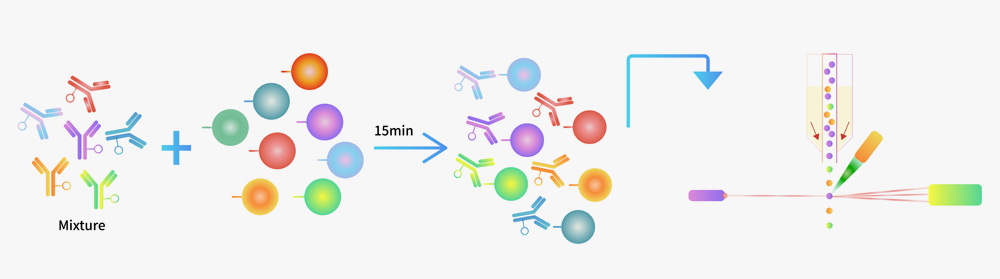
检测流程示意图
![]() 选择灵活:有荧光抗体和试剂盒可选,可灵活选择荧光抗体或试剂盒分析不同细胞;
选择灵活:有荧光抗体和试剂盒可选,可灵活选择荧光抗体或试剂盒分析不同细胞;
![]() 操作时间短:使用混合抗体组合一步染色,实验流程仅需15 min;
操作时间短:使用混合抗体组合一步染色,实验流程仅需15 min;
![]() 标准细胞确认:经标准细胞确认真实样本细胞比例准确度;
标准细胞确认:经标准细胞确认真实样本细胞比例准确度;
![]() 通量高:可一次性检测50个样本;
通量高:可一次性检测50个样本;
![]() 高特异性:Isotype Control、FMO Control及Mock细胞对照验证均无非特异性结合;
高特异性:Isotype Control、FMO Control及Mock细胞对照验证均无非特异性结合;
![]() 最优抗体组合和浓度:试剂盒包含最佳抗体和染料组合,大大提升多色荧光实验的灵敏度;
最优抗体组合和浓度:试剂盒包含最佳抗体和染料组合,大大提升多色荧光实验的灵敏度;
![]() 可靠的数据验证:经过PBMC真实样本验证;
可靠的数据验证:经过PBMC真实样本验证;
![]() 成本可控:基于自主抗体原料和荧光素偶联工艺开发,产品性能和供货能力有保障。
成本可控:基于自主抗体原料和荧光素偶联工艺开发,产品性能和供货能力有保障。
")
Flow cytometric analysis of Jurkat cells staining with APC-Labeled Monoclonal Anti-Human CD3 Antibody, Mouse IgG2a (Clone: OKT3) (Cat. No. CDE-AHFP1) at 1:50 dilution (2μL of the antibody stock solution corresponds to labeling of 1e6 cells in a final volume of 100 µL) , compared with isotype control antibody. APC signal was used to evaluate the binding activity (QC tested).
")
Flow cytometric analysis of Raji staining with PE-Labeled Monoclonal Anti-Human HLA-DR Antibody, Mouse IgG2a(L243) (Cat. No. FABm020-01) at 1:20 dilution (5 μL of the antibody stock solution corresponds to labeling of 1e6 cells in a final volume of 100 µL), compared with isotype control antibody. PE signal was used to evaluate the binding activity (QC tested).
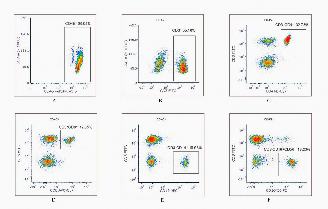
Start by setting the CD45/SSC scatter plot to identify the CD45+ lymphocyte population (A). Within the CD45+ gate, use the CD3/SSC scatter plot to identify the CD3+ T cell population (B). Next, use the CD3/CD4 scatter plot to identify the CD3+CD4+ helper T cells (Th gate) (C), and the CD3/CD8 scatter plot to identify the CD3+CD8+ cytotoxic T cells (Tc gate) (D). For B cells, use the CD3/CD19 scatter plot to identify the CD3-CD19+ population (B gate) (E). Finally, set the CD3/CD16+CD56 scatter plot to assess the CD3-CD56+CD16+ NK cell population (F).
 关注公众号
关注公众号