


 收藏产品
收藏产品 产品信息
分子别名(Synonym)
PDCD1,PD1,CD279,SLEB2
表达区间及表达系统(Source)
PE-Labeled Human PD-1, Fc,His Tag (Cat. No. PD1-HP2F2) is produced via site-specific conjugation of PE to Human PD-1, Fc,His Tag under optimal conditions with a proprietary technology. Human PD-1, Fc,His Tag is expressed from human 293 cells (HEK293). It contains AA Leu 25 - Gln 167 (Accession # Q15116-1).
Predicted N-terminus: Leu 25
Request for sequence
蛋白结构(Molecular Characterization)

This protein carries a human IgG1 Fc tag at the C-terminus, followed by a polyhistidine tag.
The protein has a calculated MW of 44.7 kDa.
偶联(Conjugate)
PE
Excitation Wavelength: 488 nm / 561 nm
Emission Wavelength: 575 nm
应用说明(Application)
Flow Cytometry (Neutralizing assay), Please note that this product is NOT compatible to streptavidin detection system.
制剂(Formulation)
Lyophilized from 0.22 μm filtered solution in PBS, 0.5% BSA, pH7.4 with trehalose as protectant.
Contact us for customized product form or formulation.
重构方法(Reconstitution)
Please see Certificate of Analysis for specific instructions.
For best performance, we strongly recommend you to follow the reconstitution protocol provided in the CoA.
存储(Storage)
For long term storage, the product should be stored at lyophilized state at -20°C or lower.
Please protect from light and avoid repeated freeze-thaw cycles.
This product is stable after storage at:
- -20°C to -70°C for 12 months in lyophilized state;
- -70°C for 3 months under sterile conditions after reconstitution.
产品数据图
活性(Bioactivity)-FACS
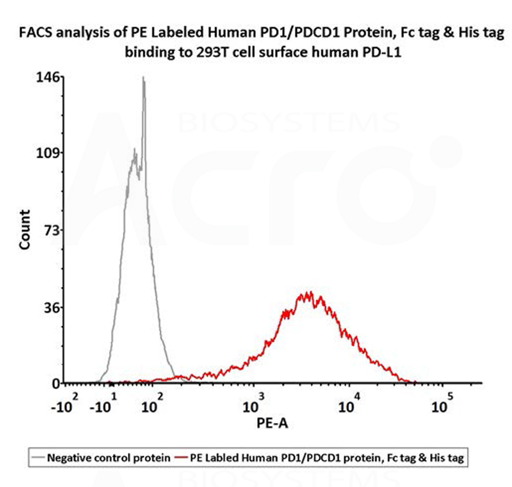
Flow Cytometry assay shows that PE-Labeled Human PD-1, Fc,His Tag (Cat. No. PD1-HP2F2) can bind to 293T cells overexpressing human PD-L1. 1 μL stock solution per million cells (QC tested).
Protocol
产品评论 发表评论

背景
Programmed cell death protein 1 (PD-1) is also known as CD279 and PDCD1, is a type I membrane protein and is a member of the extended CD28/CTLA-4 family of T cell regulators. PDCD1 is expressed on the surface of activated T cells, B cells, macrophages, myeloid cells and a subset of thymocytes. PD-1 has two ligands, PD-L1 and PD-L2, which are members of the B7 family. PD-L1 is expressed on almost all murine tumor cell lines, including PA1 myeloma, P815 mastocytoma, and B16 melanoma upon treatment with IFN-γ. PD-L2 expression is more restricted and is expressed mainly by DCs and a few tumor lines. PD1 inhibits the T-cell proliferation and production of related cytokines including IL-1, IL-4, IL-10 and IFN-γ by suppressing the activation and transduction of PI3K/AKT pathway. In addition, coligation of PD1 inhibits BCR-mediating signal by dephosphorylating key signal transducer. In vitro, treatment of anti-CD3 stimulated T cells with PD-L1-Ig results in reduced T cell proliferation and IFN-γ secretion. Monoclonal antibodies targeting PD-1 that boost the immune system are being developed for the treatment of cancer.
 靶点信息
靶点信息  数据表和文档
  联系我们
联系我们 


 项目合作
项目合作